
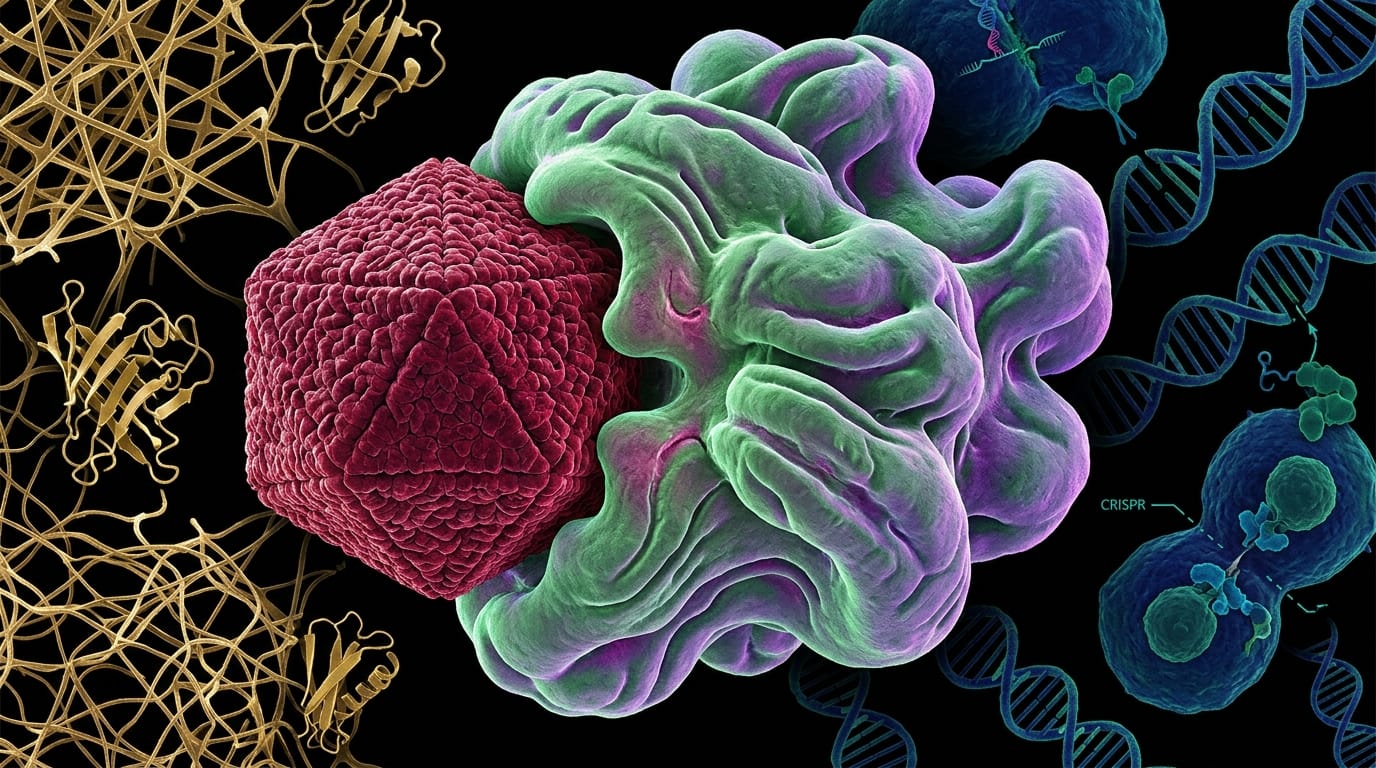
One tiny virus, the human papillomavirus or HPV, causes roughly ninety percent of all cervical cancers. It is also a culprit in many anal and oropharyngeal cancers. This virus accomplishes its devastating work by hijacking our own cellular machinery, specifically targeting a crucial guardian against tumors: a protein called p53. The idea that viruses could cause human cancer became widely accepted in the 1980s, and by 1990, scientists had already pinpointed that a particular HPV protein, known as E6, was the key player in dismantling p53.
The problem, for decades, was that we didn't fully understand how E6 did its dirty work. E6 doesn't act alone; it needs a human helper protein, called E6AP. Together, these two proteins—one viral, one human—form a potent team, and they recruit p53 into what scientists refer to as the HPV16 E6-E6AP-p53 degradation complex. This complex acts like a molecular wrecking crew, marking p53 for destruction. Knowing this complex was the enemy, researchers spent years trying to design drugs to stop it. Yet, these efforts largely failed. A major reason for this was a missing piece of the puzzle: a high-resolution map of the complex itself. Earlier techniques, like nuclear magnetic resonance, struggled to fully resolve the structure of the E6 protein, which behaves poorly on its own.
Now, a powerful imaging technique has changed everything. Think of it this way: instead of trying to understand a complex machine by looking at its blueprints or blurry photos, we can now see a detailed, three-dimensional model of it, frozen in action. This is what single-particle cryo-electron microscopy (cryo-EM) allows. Using cryo-EM, Pachano, Tomas, Leng, Dugied, Tribble, Loubiere, and their colleagues achieved an atomic-level view of the entire HPV16 E6-E6AP-p53 degradation complex. This incredibly detailed cryoEM structure, resolving the complex down to roughly three atoms in width, finally provides the comprehensive map scientists have desperately needed.
The key thing to understand here is what this detailed map revealed about the interaction between the viral E6 protein and its human accomplice, E6AP. The structure shows that these two proteins don't just lightly touch; they have a surprisingly large and extensive contact area, what researchers call a protein-protein interaction interface. Imagine two hands gripping each other tightly over a broad surface rather than just by the fingertips. This expansive connection explains something crucial: the incredibly strong attraction between HPV16 E6 and E6AP. Scientists measured this powerful bond as having a picomolar binding affinity, meaning these two proteins stick together with extreme tenacity. This kind of intense binding makes them incredibly difficult to pull apart.
With that foundation established, we can now see precisely how this stable, integrated complex leads to the downfall of p53. The structure helps us understand how the viral E6 protein, with E6AP's assistance, effectively tags p53 for removal. This tagging is the critical step in p53 degradation, leading to its breakdown and clearing the way for uncontrolled cell growth—the hallmark of cancer. Previous research had only a limited understanding of how E6AP and E6 truly connected beyond a small, known docking region. This new comprehensive view gives us the full picture.
The structural understanding offers a profound reevaluation of past drug development strategies. For more than two decades, the HPV E6-E6AP-p53 complex has been a prime target for drug development. But the limited success in creating effective therapies might now be explained by this newly revealed intricate stability and the extensive protein-protein interactions. Without seeing this strong, widespread handshake between the viral and human proteins, previous drug efforts might have been akin to trying to pry apart those tightly clasped hands with imprecise tools. The research also reminds us that not every part of E6AP could be resolved due to its flexibility, hinting at the dynamic nature of these molecular machines and the challenges in targeting them.
This detailed molecular map unlocks a critical path forward: rational drug design. Instead of searching blindly for compounds that might disrupt the complex, scientists can now use the high-resolution structure to identify specific pockets or interaction interfaces. They can design molecules that act like perfectly fitted wrenches, precisely jamming the gears of this cancer-promoting machinery. This approach moves beyond trial-and-error, paving the way for the first generation of truly targeted therapies for HPV-driven cervical, anal, and oropharyngeal cancers. The implications are profound, offering new hope in combating the global burden of these devastating diseases. ◆
Sourced from: "Systematic discovery of pathogen effector functions across human pathogens and pathways." by Pachano, Tomas et al.