
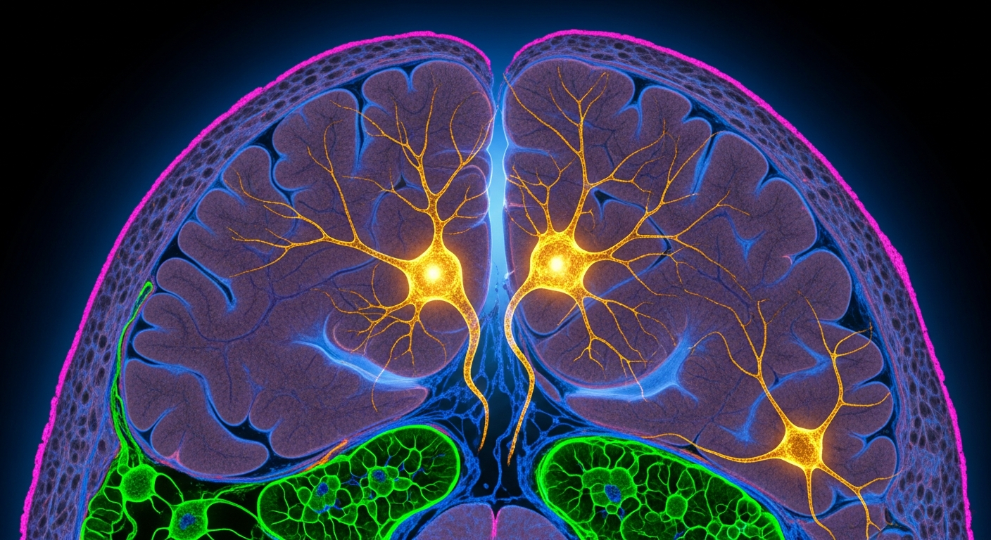
Imagine trying to read the specific chemical instructions firing within individual brain cells, not just anywhere, but in the tiny, intricate regions of the human hypothalamus, long after the brain has been preserved. The hypothalamus acts as the body's central thermostat for energy, meticulously balancing how much we eat with how much energy we burn. For years, scientists have understood that two key types of neurons in this area, Agouti-related protein (AgRP) neurons and proopiomelanocortin (POMC) neurons, play opposing but critical roles in this balancing act. AgRP neurons essentially hit the "on switch for appetite and tell the body to store energy, while POMC neurons act as the "off switch, suppressing feeding and encouraging energy expenditure. Yet, mapping the exact genetic blueprint — the unique set of active genes — within these specific human cells has been a significant challenge. The difficulty lies in how brain tissue is typically preserved and analyzed. Our brains are incredibly complex structures, and singling out specific, tiny cell populations to understand their internal molecular workings requires extremely refined tools. The goal isn't just to see that these neurons exist, but to understand what they are doing at a fundamental level by looking at their active genes, or transcripts. The insights gained from such detailed mapping could illuminate the molecular mechanisms that govern our body’s energy balance, offering clues for new approaches to weight management.
This is where a new technique, pioneered by Katalin Skrapits, Dalma Biri, and Erik Hrabovszky in their research, enters the picture. They developed and verified a method called IHC/LCM-Seq, designed to precisely profile the active genes within specific neuronal cell types. This method is particularly clever because it works with human brain tissue that has been fixed with formaldehyde, a common preservation technique that makes the tissue durable but historically difficult for detailed genetic analysis.
The IHC/LCM-Seq method involves two main steps. First, immunohistochemistry (IHC) helps researchers identify and visually tag the exact types of neurons they want to study — in this case, the AgRP and POMC neurons — within thin slices of brain tissue. Once these specific cells are marked, laser capture microdissection (LCM) uses a microscopic laser to literally cut out and collect only those targeted neurons. Finally, the "Seq part refers to sequencing the genetic material from these isolated cells, allowing scientists to create a comprehensive list of all the active genes. This multi-step process means researchers can now isolate and analyze the genetic activity of very specific cell populations from archived human brain samples, providing a flexible way to study cell types in postmortem human brain tissue.
Using this new method, Skrapits, Biri, and Hrabovszky applied IHC/LCM-Seq to male human hypothalami. They found that both the agouti-related protein (AgRP) neurons and the proopiomelanocortin (POMC) neurons contained between 14,000 and 16,000 active genes. This number — tens of thousands of active genes in each type of neuron — indicates a rich and complex molecular profile, distinct for each cell type. The discovery of these specific molecular signatures in AgRP and POMC neurons offers new insights into how these cells carry out their unique tasks of either driving appetite and energy storage, or suppressing feeding and promoting energy use. AgRP neurons, for example, showed active genes related to inflammation, metabolic processes, and growth, which aligns with their role in increasing appetite. POMC neurons, on the other hand, displayed active genes for receptors involved in reproductive hormones, a signaling molecule called glucagon-like peptide, and endocannabinoids, all of which fit with their function in reducing food intake and boosting energy expenditure.
It is important to remember, however, that this work, as presented in the manuscript, is unedited. It will undergo further review and editing before it is formally published. This means that, while the method itself is a significant development and the findings are intriguing, the precise details and interpretations may yet be refined. The study focused specifically on male human hypothalami, which provides valuable human data, but also means that any conclusions about these neurons might not directly apply to female brains or to how these systems function in a living person. The data reveal the potential for distinct molecular profiles, but more work is needed to fully understand their dynamic roles in living individuals and across different populations.
What these findings actually give us is a much clearer, high-resolution map of the genetic landscape inside the human brain's energy control centers. This detailed picture could help researchers identify specific biological molecules or pathways within these neurons that might serve as new targets for weight-management strategies. Instead of broad interventions, the hope is to develop more precise approaches that specifically influence the activity of AgRP or POMC neurons. The careful mapping of these cellular codes is a crucial first step in a long, complex journey toward developing more effective ways to help the body maintain its delicate energy balance. ◆